The rise of precision medicine has brought with it unprecedented breakthroughs in biologic drug development, particularly related to immunotherapy. These novel compounds offer the exciting prospect of treating patients with targeted therapies aimed at their disease’s specific characteristics and genetics. However, with biologically-based therapeutics comes a greater need for information that supports aspects like target selection, dosing determination, and administration schedules, including an understanding of the drug’s pharmacokinetics (PK), pharmacodynamics (PD), and receptor occupancy (RO).
RO assays qualitatively and/or quantitatively measure how a biotherapeutic is binding to its cellular target, and they have become a critical component of the biomarker portfolio for protein‐based therapeutic agents targeting cellular antigens, such as those directed at immune conditions and cancer. These assays can add value in every stage of the development lifecycle, from helping to model starting doses for early Phase I trials, to determining the efficacious dosing range in Phase II, to helping define population PK characteristics of the biotherapeutic and covariates that could impact its mechanism of action in Phase III. As such, there are a variety of approaches available to perform these assays.
So what makes flow cytometry the platform of choice for receptor occupancy studies? This versatile method affords the ability to measure individual targets/receptors using fluorescent tags in specific cell populations, making it ideally suited to RO measurements.
The Importance of RO Assays by Flow Cytometry
RO assays on the flow cytometric platform (also called FACS analysis) can be designed for different levels of complexity, from simply measuring the number of cell surface receptors bound by an anti-receptor therapeutic agent to more intricate measurements of receptor internalization and shedding events once the receptor engages the therapeutic. The platform can measure distinct subsets in complex heterogeneous populations, including:
- Free receptor determinationusing a competitive antibody or fluorescently labeled biotherapeutic agent (often employed with monoclonal-based therapeutics) to determine the number of receptors not occupied by the biotherapeutic.
- Total receptor detection using noncompetitive antibodies to detect all expressed receptor targets, whether the biotherapeutic is present or not, to measure the total expression on the surface of the cell. Using free and total receptor assays in combination can support assessment of achieved saturation by determining the ratio of unoccupied sites to total sites available at any given time during dosing.
- Bound receptor measurement most often using a fluorescently labeled antidrug antibody to detect the biotherapeutic agent bound to the target receptors; although if the drug is a monoclonal antibody (mAb), a detection antibody that does not competitively inhibit the drug-target binding may be used as the drug detection reagent.
- Receptor modulation assessment for monitoring the effect on the modulation of the target receptor or other functional effect that results from biotherapeutic binding, such as inhibiting internalization of the receptor. Free and total RO assays can again be used in combination here to assess if receptor modulation occurs in response to treatment over time.
Measuring RO throughout development is particularly useful when working with mAbs, as most in use have long biological half-lives and an understanding of their binding characteristics is critical to therapeutic decision-making. Improper dosing can lead to severe adverse effects, and therefore the European Medicines Agency (EMA) recommends selecting a starting dose (retrieved from non-clinical pharmacology data) that would result in a Minimum Anticipated Biologic Effect Level (MABEL) for therapeutics for which a potential risk of side effects has been identified. Non-clinical pharmacology PK/PD modeling and the use of RO assays are fundamental tools for correctly determining the MABEL to guide the pharmacologically active dose selection and to anticipate therapeutic dose range (ATD) in humans.
Flow Cytometric Measurement of RO in Clinical Trials
It is important to note that when using RO assays in clinical trials, day-to-day variability must be taken into account. At this point, in addition to measuring the frequency of target cell populations, the fluorescence output of labeled reagents needs to also be assessed and controlled. Beads can be used to establish target ranges and normalize the flow cytometer before the RO assays are run, ensuring comparability in the data collected over time.
Parter with BioAgilytix | In-House Flow Cytometry Platform
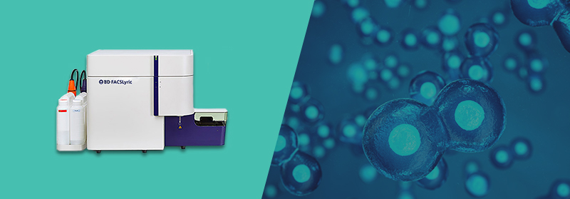
RO assays are just one of several ideal applications for the flow cytometry platform. In our upcoming blog series, we’ll be discussing the value of flow for immunophenotyping and for immunogenicity assessment using cell-based assays. In the meantime, you can find more information on our own flow cytometry platforms, including BD FACSLyric™, here or schedule a discussion with our scientists to talk through your RO assay requirements and how we can support them.
