If your goal is to attain swift and simple protein quantification, the Bradford assay is the way to go. Let’s find out more about what makes the Bradford assay so effective and relevant over 40 years after its inception.
What is Bradford Assay?
The Bradford protein assay, named after its developer Marion M. Bradford, is specifically used to calculate the concentration of total protein in a sample or solution. There are three standard methods to calculate and measure protein concentration and those are the bicinchoninic acid assay (BCA assay), absorbance at 280 nm and the Bradford Assay.
Bradford assays are conducted at room temperature, require no special equipment, and are compatible with most solvents, salts and buffers. Bradford protein assays are Coomassie dye-binding assays that result in the sample’s color-changing from brownish-green to blue during the process. This color-changing reaction is dependent on the amino acid composition of the measured proteins.
How the Bradford Protein Assay Works?
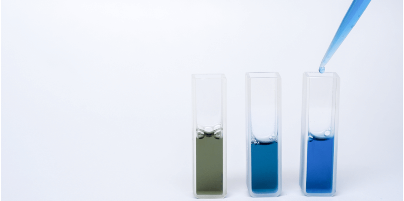
Bradford protein assays are Coomassie dye-binding assays. When the Bradford reagent binds to proteins it results in the sample’s color-changing from brownish-green to blue. This color-changing reaction is dependent on the amino acid composition of the measured proteins, with the absorbance maximum moving from 470 to 595 nm.
There are three forms of Coomassie Brilliant Blue G-250 dye: Anionic (blue), neutral (green), and cationic (red). The Bradford assay is brilliant because it allows you to measure the color intensity and observe your findings with the naked eye. The color of the Bradford reagent should be brown or reddish-brown with lambda max 470 nm and could plausibly be a dishwasher-green color without cause for alarm. However, if you haven’t yet added a protein, there is no reason it should be blue.
The National Institutes of Health (NIH) explained it in this context, “The determination of microgram quantities of protein in the Bradford Coomassie brilliant blue assay is accomplished by measurement of absorbance at 590 nm. This most common assay enables rapid and simple protein quantification in cell lysates, cellular fractions, or recombinant protein samples, for the purpose of normalization of biochemical measurements.”
Calculating Protein Concentration Utilizing Bradford Assay
Traditionally, the preferred method for calculating protein concentration using the Bradford assay is a standard curve generated from known protein standards. Because it is difficult to select a protein standard that is complementary to the sample being analyzed, it has become scientifically acceptable to use common proteins like bovine serum albumin (BSA) and gamma globulin as standards.
The only drawback of using BSA or the bovine γ-globulin (IgG) as reference proteins is that there is a considerable variation between proteins and yields results that are only an approximation of protein concentration.
You can use either a spectrophotometer or a microplate reader to calculate the absorbance at 595 nm. The precise protein concentration of the sample is determined by the interpolation from a standard curve made by measuring the absorbance of a dilution series of protein standards of known concentrations within the linear response range of the Bradford protein assay.
Calculation Procedures (Credit)
Materials –
- BSA standard solution (0.1 µg/µl)
- Bradford solution – Dissolve 100 mg Coomassie Brilliant Blue G-250 in 50 ml 95% ethanol. Add 100 ml of 85% phosphoric acid while stirring continuously. When the dye has dissolved, dilute to 1 l in H2O. Filter to remove residual precipitate and store at 4 °C in a dark bottle.
- Spectrophotometer (Tecan)
- Whatman #1 paper (Whatman)
Preparation and Process –
- Prepare protein standards according to the following scheme
- Prepare a 1:10 dilution of your protein sample (=SPL 10)
- Prepare a dilution series of SPL10 according to the following scheme. For protein concentrations between 5-20 mg/ml it is sufficient to prepare SPL40-SPL200
- Transfer 10µl duplicates of each standard and sample into a 96-well plate and add 180µl Bradford reagent
- Incubate 10min at room temperature and measure absorbance at 595nm
- Prepare a calibration curve by plotting absorbance versus protein amount in µg
- Determine the amount of protein in your samples using the trend line
The Premiere Cell-Based Assay & Immunoassay Lab at BioAgilytix
BioAgilytix is the world’s leading large-molecule bioassay lab. We provide the specialized large molecule insight and proven GLP / GMP knowledge needed to support a full range of cell-based assay requirements. Our veteran team possesses expertise with primary cells and transformed cell lines. Working with our customers, we are able to develop and validate custom cell-based assays in line with all relevant regulatory guidelines and recommendations.
BioAgilytix is the clear choice for cell-based assay and immunoassay development and analysis. We provide accuracy, reliability and world-class expertise. Contact BioAgilytix today for more information on our wide range of bioanalysis services.