Flow Cytometry – What’s Old is New Again
Analytical testing of therapeutic compounds often requires data at the single-cell level. Flow cytometry is a versatile technology that can analyze multiple characteristics of cells and provides a wealth of data from clinical samples. A few examples include:
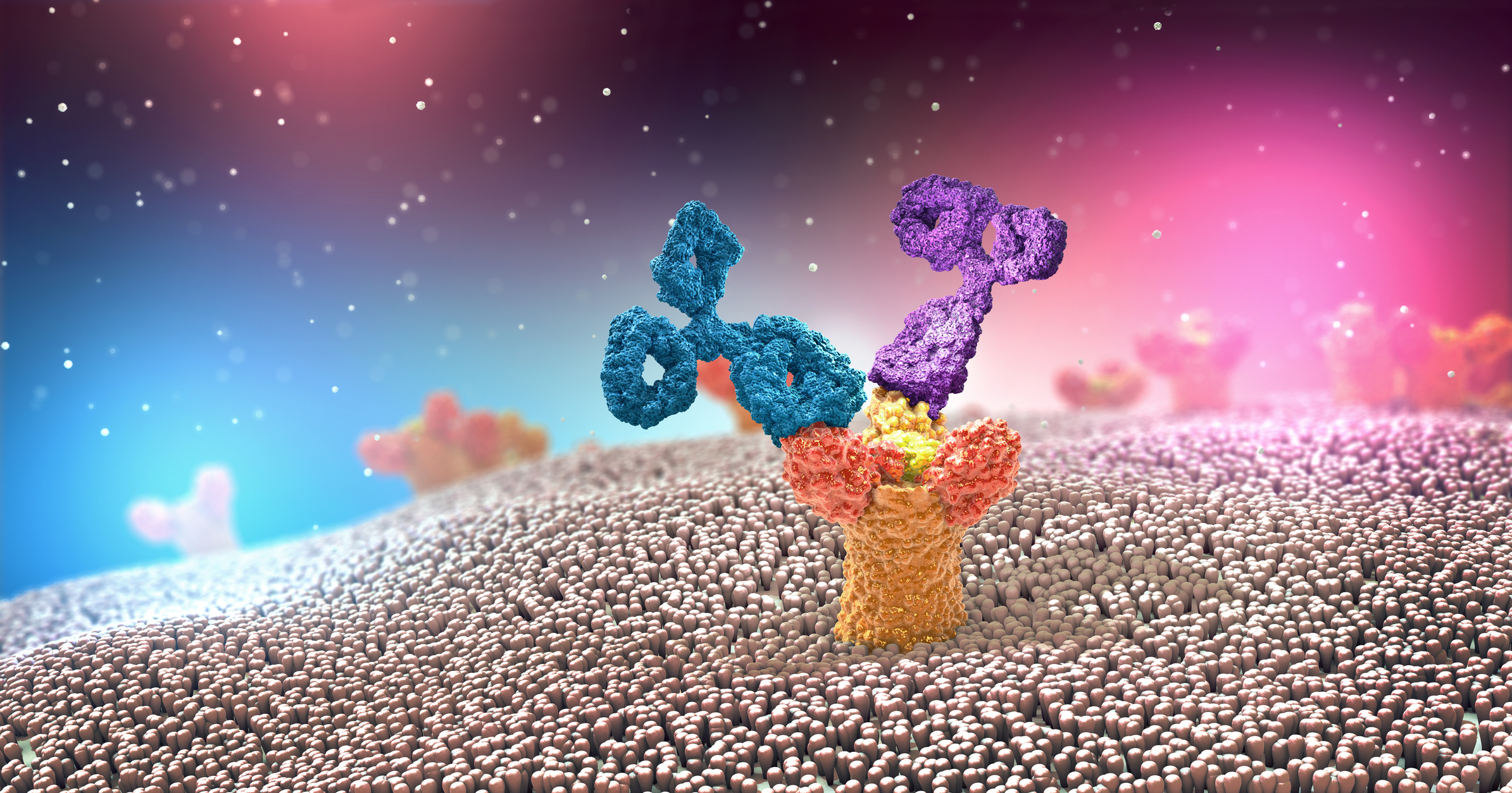
- For antibody-based therapeutics, it is often necessary to understand the binding of a drug to a cell surface target, known as a receptor occupancy assay.
- For immune modulating drugs, the immune cell profile of a patient may need to be established with an assay to enumerate the specific immune cell subsets within blood samples.
- For cell therapies, distinguishing and quantitating the therapeutic from the patient’s own cells is necessary to capture the persistence of the drug product as part of the pharmacokinetic profile.
In this post, the first in a three-part series, we’ll explore how flow cytometry came about and how the technology has advanced over the years.
A Brief History of Flow Cytometry
Flow cytometry is a robust and well-established bioanalytical platform that was first introduced in the 1950s. Although the current methodologies and equipment have become much more complex thanks to technological advances, the original principle remains the same: Cells can be made to flow through a narrow opening in a single-cell stream and then interrogated mid-flow to provide information such as size, composition, and phenotype
Technical Advancements
More modern equipment involving lasers and microfluidics has been developed to enable detailed information and multiplexed read-outs. A sophisticated array of lasers, optics, fluidics, and electronic detectors can measure light scatter and/or fluorescence emission from cells. Additional techniques including acoustic focusing, cell sorting, and spectral flow analyzers have been introduced in more recent years, expanding on the technical options within the field.
Through the use of fluorescent antibodies and dyes, individual markers or phenotypic aspects on cells can be labeled and analyzed. Flow assays can be designed to identify specific subpopulations of cells from a larger, mixed group based on identifiers such as surface markers, size, and granularity. In addition to proteins expressed on the surface of cells, intracellular proteins can be labeled by fixing and permeabilizing the cells prior to labeling. DNA dyes and viability stains can be used to measure cell cycle, proliferation, and apoptosis.
BioAgilytix Uses Flow Cytometry in Immunoassays
BioAgilytix performs routine testing using the CytoFLEX LX, BD FACSLyric™, BD LSRFortessa™ X-20, and BD FACSCanto™ II flow cytometry platforms as part of our premier immunoassay capabilities. We utilize this equipment to support both preclinical and clinical work in non-regulated and regulated environments for a variety of assay end-points including, but not limited to:
- Immunophenotyping
- Intracellular cytokine staining
- Proliferation
- Apoptosis
- Reporter cell signaling
- Biomarker analysis
- Cell therapy measurements
Our experienced scientific team can work directly with you to design and develop specialized assays to fit your flow cytometry needs.
Don’t Miss the Multiparameter Flow Cytometry Webinar
Register for our upcoming webinar, “Multiparameter flow cytometry as a powerful tool in drug development” to learn more about the role of flow cytometry in drug development as well as other real-world applications.
References: